Ligament reconstruction is most commonly undertaken for symptomatic instability (giving way) of the knee after anterior cruciate ligament (ACL) rupture. This usually occurs after an injury to the knee and is commonly sustained during sporting activity. Using the patient’s own tendon tissue (referred to as autograft), a new ACL is fashioned and implanted in the knee during an arthroscopic (minimally invasive) operation. Substantial rehabilitation under the supervision of a physiotherapist is then required until the new ligament is fully healed. Return to normal activities will depend on the patient’s requirements but normal walking is possible within weeks of the procedure. Return to strenuous sports such as football or squash usually takes six to nine months but non-twisting exercise can usually be started sooner. Fortunately, surgical reconstruction of the other knee ligaments (posterior cruciate and collateral ligaments) is seldom required unless a major injury has been sustained.
Magnetic resonance imaging (MRI) scans showing a normal, intact anterior cruciate ligament (ACL) on the left image and a torn ACL on the right image. The ACL is indicated in both images by an arrow and is outlined in red.
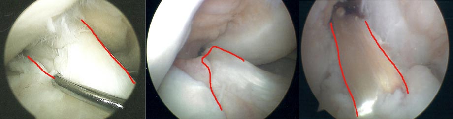
Arthroscopic views inside the knee showing the anterior cruciate ligament (ACL) outlined in red. From left to right: normal ACL, torn ACL and reconstructed ACL. The metal device on the left image is a probe used to check the ACL.